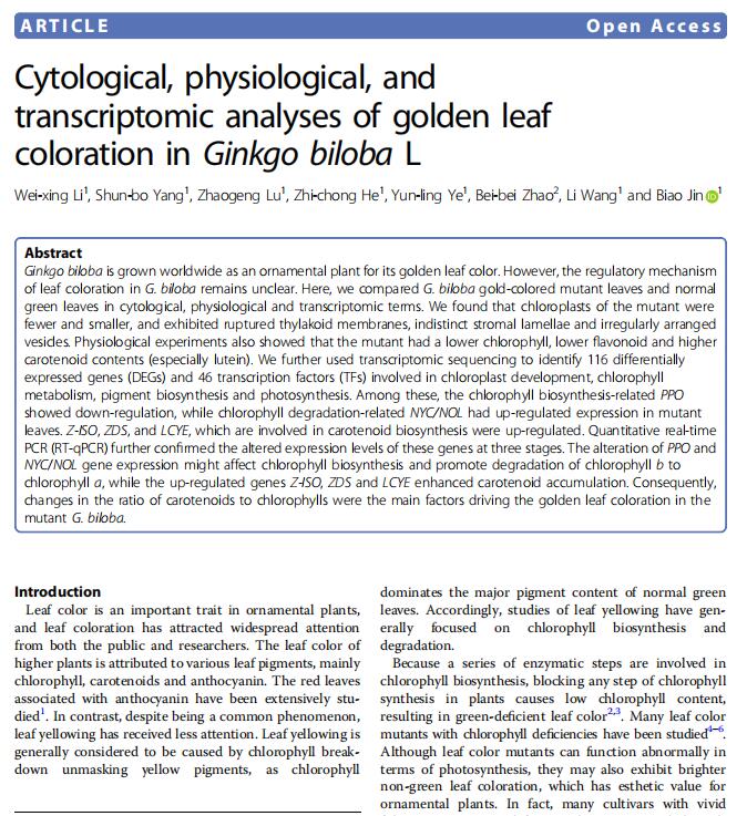
【文獻標題】 Cytological, physiological, and transcriptomic analyses of golden leaf coloration in Ginkgo biloba L
【作者】 Wei-xing Li, Shun-bo Yang, Zhaogeng Lu,et.al
【作者單位】 揚州大學
【文獻中引用產品】
有機氯ELISA試劑盒(有機氯農藥殘留快速檢測ELISA檢測試劑盒)
【DOI】doi.org/10.1038/s41438-018-0015-4
【影響因子(IF)】 4.21
【出版期刊】 《Horticulture Research》
【產品原文引用】
Materials and methods
Plant material
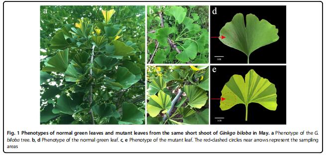
A healthy adult G. biloba tree bearing both normal green leaves and golden–green striped leaves was used in this study, after growing under natural conditions in the Ginkgo nursery of Yangzhou University (32°39′ N, 119°42′E). Normal green leaves and golden–green striped leaves borne on the same short shoot of this tree were collected from May to July in 2015 and 2016 (Fig. 1a–c). For cytological, physiological and RNA-Seq experiments,yellow parts of the golden–green striped leaves (mutant leaves) and the green leaves (normal leaves) were sampled separately in May. For RT-qPCR, the mutant leaves and green leaves from three stages (May to July) were used. All of the samples were collected from the identical spatial sections of leaves (Fig. 1d, e), and immediately frozen in liquid nitrogen, and stored at −80 °C until use.
Measurements of chlorophyll, carotenoid, and flavonoid contents Approximately 0.1 g of leaves from normal green leaves and the golden parts of mutant leaves (sampled in May) were cut into pieces and submerged in 80% acetone Fig. 1 Phenotypes of normal green leaves and mutant leaves from the same short shoot of Ginkgo biloba in May. a Phenotype of the G.biloba tree. b, d Phenotype of the normal green leaf. c, e Phenotype of the mutant leaf. The red-dashed circles near arrows represent the sampling areas Li et al. Horticulture Research (2018) 5:12 Page 2 of 14 overnight to extract the chlorophyll. Next, the extract was measured spectrophotometrically at 665, 649, and 470 nm with a UV-1800 spectrophotometer (DU-800; Beckman Coulter, Brea, CA, USA). Soil-plant analysis development (SPAD) values were measured in situ on each type of leaf using a Chlorophyll Meter SPAD-502Plus (Konica Minolta, Osaka, Japan). To measure the contents of chlorophyll intermediaries, leaves were homogenized in nine volumes of 0.01 M phosphate-buffered saline, mixed on ice, and centrifuged (30 min at 2500 g). The supernatant was assayed separately with an ELISA kit (HengYuan Biological Technology Co., Ltd, Shanghai, China). Carotenoid components were measured using highperformance liquid chromatography, and total carotenoid and flavonoid contents were determined using a UV-1800 spectrophotometer. Three biological replicates were evaluated for each sample. The data thus obtained were analyzed using SPSS software (ver. 17.0; SPSS Inc.,Chicago, IL, USA)


完整版PDF文獻請咨詢在線客服或者聯系我司業務員!
更多公司福利請關注“恒遠生物”VX公眾號!
